My Rambling Thoughts
  • Home
  • Blog

Vaccines: why they help everybody (not just the vaccinated) but only when most people get them

11/1/2017

1 Comment

 
PictureFrom: http://www.thevaccinemom.com/2015/02/vaccine-shedding-should-you-really-be-concerned/
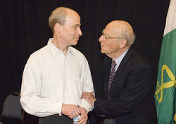
Vaccination has led to disappearance of diseases that killed thousands less than a century ago

There are many diseases we have never heard of anyone suffering from because there are effective vaccines against them. Some of these diseases are on their way to being (or have been) almost or completely eradicated - diphtheria, polio, smallpox, whooping cough (pertussis), measles, mumps, rubella. Because of effective vaccines available to all, in developed areas of the world disease incidence is almost negligible. In contrast, other regions have considerable numbers of cases every year, some resulting in death. According to UNICEF, almost one third of deaths among children under 5 years of age are vaccine-preventable. In 2015 about one in five infants (that is 20% of all infants or 19.4 million children) did not get important vaccines- most of them among the poorest and more vulnerable populations. In these areas, the need for vaccination is obvious, but in developed countries some people may have a hard time understanding this need. Pediatricians and schools requesting that babies and children follow the recommended vaccination schedule make it all happen smoothly most of the time. Unfortunately, some conspiracy theories supporters have negatively affected vaccination rates in recent years.

Why do we need to get vaccinated if the chances of getting sick are almost zero? Because if we don't these chances increase, and not just for  us but for others as well (see below), and diseases eventually come back. Here are some examples when a reduction in vaccination rates led to reintroduction of diseases to much higher levels including deaths:
1) Diphtheria in the former Soviet Union in the 1990s: low children vaccination and a lack of booster vaccinations for adults resulted in nearly 50,000 cases (from 839 cases in 1989) and 1,700 deaths in 1994. 
2) Lower use of pertussis (whooping cough) vaccine because of fear about the vaccine in:
2.1) The UK: an epidemic of more than 100,000 cases and 36 deaths by 1978
2.2) Japan: vaccination went from 70% to 20%-40% leading to a dramatic rise from 393 cases and no deaths in 1974 to 13,000 cases and 41 deaths in 1979
2.3) Sweden: incidence increased from 700 cases per 100,000 children 0-6 years of age  in 1981 to 3,200 in 1985.


PictureFrom http://www.mydr.com.au/travel-health/vaccination-and-antibodies
How do vaccines work and what do they contain?

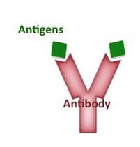
Vaccines are designed and engineered using parts or the whole microorganism that causes the disease (virus or bacteria) after it has been “weakened”: they are killed or inactivated in specific ways that leave them unable to replicate or cause disease. Our immune system “sees” these “antigens” thanks to vaccination and produces antibodies that specifically recognize these antigens. Some immune memory "B" cells remain after this response to the vaccine ends and the antibodies disappear. The immune system later “remembers” these antigens when exposed to the live pathogenic agent that can cause disease and activates the memory B cells resulting in a much stronger and lost lasting response producing tons of antibodies. As vaccines usually result in life-long immunity, whenever we are exposed to the pathogen our immune system will mount a strong immune response which will control the infection regardless of the time passed since vaccination. Even if vaccine protection is not 100%, it will provide some protection so we do not get the full blown disease and symptoms that we would if we were not vaccinated.

The effect of a vaccine can be enhanced by the addition of "adjuvants" (from Latin: helpers) that go along with the antigens from the virus or bacteria and improve the quality and/or quantity of the immune response, although their exact action mechanisms are not well understood. Aluminum and oil-in-water are common adjuvants used in vaccines. Aluminum salts are used in DTaP, pneumococcal and hepatitis B vaccines in the US. All adjuvants (as parts of vaccines) are tested in long clinical trials to evaluate safety and purity before approval by the FDA in the US, including aluminum vaccines that have been used for over 60 years and only very rarely result in severe local reactions. We get regularly exposed to aluminum from food and water also. 

The past use of the mercury-based preservative called thimerosal raised concerns about possible toxicity although no adverse effects occurred except for  expected minor irritation at the injection site. Since 2001, except for some flu vaccines, no vaccines used in young children contain thimerosal.

Picture
Herd Immunity and diseases making a comeback due to low vaccination rates

Only smallpox has been eliminated globally. Polio is on its way, but a few countries still have cases. More than 350,000 cases of measles occurred in 2011 around the world, with 90% of US measles cases imported from another country. In 2013 however, several measles outbreaks occurred in the US including Texas and New York City in low vaccination populations. These outbreaks due to reduced vaccination rates threaten the progress made towards disease eradication.

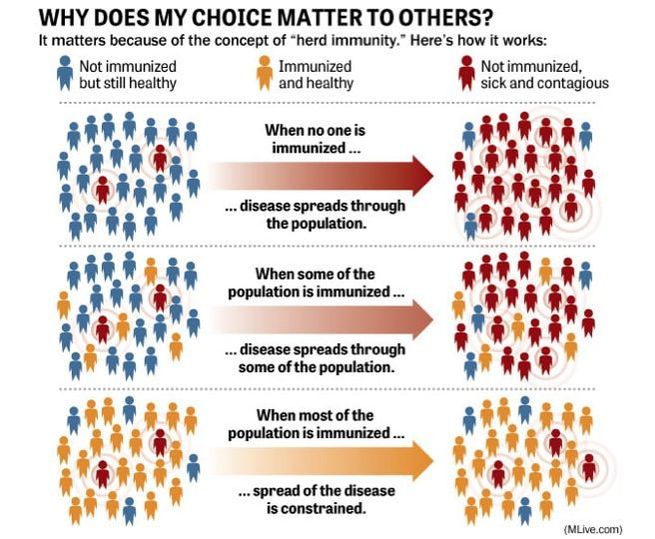
Most public health vaccination programs aim at 100% vaccination coverage. But there’s always a proportion of the population that isn’t vaccinated- babies too young to be vaccinated and people with certain conditions such as those who are immunocompromised (HIV infected or people receiving chemotherapy). However, if enough people get vaccinated among specific populations, the non-vaccinated get protection from the so-called herd immunity which basically blocks spreading of the infection to others even when some non-vaccinated get sick. The minimum proportion of vaccinated people necessary for herd immunity (which depends on several factors) is about 90-95%. How this works is illustrated on the figure below.
Picture
From: http://media.mlive.com/mlive_statewide_river_candidates/photo/herd-immunityjpg-e2a094c9040c0789.jpg


1 Comment

Get to know your vitamins

10/20/2017

8 Comments

 
Vitamins are natural organic (carbon-containing) substances that can be found in food (and in the very popular “supplements” that many people take in probably much higher amounts that they may need to) and that our bodies require for a variety of processes that occur inside us. They are also known as micronutrients, they are needed in small amounts and they are also small in structure compared to proteins, fats and carbohydrates that need to be broken down in order to be used, whereas vitamins do not. They are also “essential” nutrients, meaning they are essential for us to get from food as our bodies can not make them on their own except for vitamin D, which the body can make when exposed to sunlight- not very frequent event for most people in the winter. Calcium, which is absorbed in the small intestines in a process that needs vitamin D, is a common supplement that is taken with vitamin D especially for women in and after menopause to prevent osteoporosis. Vitamin K2 (a type of vitamin K) is made by some of our good gut bacteria, but we don't get much from this source.

Both vitamin deficiencies (much lower amounts than our bodies need) and excess (higher than the maximum our bodies need/tolerate) may lead to adverse symptoms for some vitamins. For example, vitamin A deficiency may result in night blindness first leading to blindness later on, and tissues like eyes and skin may become dry and damaged while infections are more likely due to the immune system not working properly especially in infants and children, whose growth and development may be slowed. However this deficiency can be reversed by taking high vitamin A doses for several days. On the other hand, excess vitamin A may result in hair loss, cracked lips, dry skin, weakened bones, headaches, and increased pressure in the brain, although these effects requires intake of really high doses and are reversed by stopping vitamin A intake.
Picture
Picture
From: http://www.stylecraze.com/articles/3-vital-vitamins-for-hair-growth/#gref

There are a total of 13 vitamins, which can be divided in two groups: those soluble in water, and those soluble in fat. All B vitamins and vitamin C are water-soluble and our bodies can’t store them. They leave us quickly via our kidneys and then in our urine, so we need to provide our bodies with them often, ideally every day. In contrast, vitamins A, D, E, and K are fat-soluble, absorbed in our intestines in fatty forms and easily stored in our bodies in the liver and fatty tissues for long periods of time (except vitamin K). This is why toxicity may occur if too much is taken of a fat-soluble vitamin, especially vitamin A or D.

All vitamins are absorbed in the small intestine, and then transported to specific tissues in the bloodstream; fat-soluble vitamins need to be transported first by the lymphatic system after intestinal absorption to the blood. At the right destination, vitamins help "reactions" occur, several carried out by enzymes that need a little help from "coenzymes" which some of these vitamins act as. Vitamins C and E are well known as "antioxidants", meaning they protect our cells from excessive accumulation of "free radicals" that may cause "oxidative stress" and result in an increased risk for cardiovascular disease, cancer and other diseases.

For a fun and informative 5min TED Ed video on how we absorb vitamins, how they are transported and what they help us with, watch: https://www.youtube.com/watch?v=ISZLTJH5lYg

                                     Water Soluble Vitamins


Picture
                                             Fat Soluble Vitamins
Picture
Information for tables above came mostly from Harvard Medical School at: https://www.health.harvard.edu/staying-healthy/listing_of_vitamins

Picture
If you eat fortified cereals for breakfast (cereals with added vitamins and minerals) you may be getting more than the daily amount of vitamins recommended, especially children, and even more so if you are taking vitamin supplements, eating fortified snack bars, etc. The daily values shown in cereal boxes (that come from the US FDA) are usually the ones for adults- children need less; some cereals show both amounts.

If we have access to good food rich in vitamins, we don’t really need to supplement our regular intake with extra vitamins, but there may be situations/places in which taking specific vitamin supplements is recommended (for all or some of them) if the food is not rich in these, or you have a diet that restricts food with certain vitamins. Keep in mind that man-made vitamin supplements usually contain the full recommended daily amount of the different vitamins, which we are taking in addition to what we are ingesting with our food, and don't forget to count the fortified food you eat that contains added vitamins (milk, cereals, flour). As I am a petite person, I feel that for things like supplements or pain killers I am probably taking way more than I need (compared to someone twice my size/weight for example) so I may take less than the recommended adult dose sometimes, and/or less frequently- disclaimer here: this is just my approach; I am not the MD kind of doctor so my advice is not that of a clinician.
8 Comments

why your circadian rHythms are important for your health

10/7/2017

1 Comment

 
In honor of the Nobel Prize in Physiology or Medicine 2017 given to Michael Young, Michael Rosbash and Jeffrey Hall for revealing molecular mechanisms controlling the circadian rhythm, this post is dedicated to this topic.
 
Circadian rhythm or clock refers to a complex mechanism underlying the capacity of living organisms (plants and animals including us) to adjust several of our body processes to the sunlight during the day and darkness at night in 24h (solar) oscillating cycles. This figure taken from the press release of the 2017 Nobel prize awarded this past October 2nd shows how the circadian clock helps us adapt our physiology to the different phases of the day and regulates sleep patterns, feeding behavior, hormone release, blood pressure, and body temperature.
Picture
“Molecularly” speaking, this clock is regulated by specific genes in different cells, that get activated and transcribed into RNA, which leads to protein production and accumulation within cells. Once a certain level is achieved, this acts as a “feedback loop” turning off expression of the genes that were activated before. The cycle takes 24h total to begin again. Several genes have been identified thanks to many years of research, mostly in Drosophila (fruit fly) and mice as the animal model for mammals. Although the central “clock” is located in the suprachiasmatic nucleus (SCN) in the hypothalamus in mammals, formed by neurons that act based on sensing light (“photic”) input from the retina, circadian rhythm clocks are also present in most peripheral tissues and cells. Food schedule also has an important impact on this clock. Organs such as liver, pancreas, adrenal gland, spleen, thymus, and heart each have their own rythm, as well as fatty cells. The hormones insulin, cortisol and melatonin are secreted (produced and released to act on target organs/tissues/cells) in a circadian-dependent manner.
Picture
From:
https://www.intechopen.com/books/molecular-mechanisms-of-the-aging-process-and-rejuvenation/circadian-clock-gene-regulation-in-aging-and-drug-discovery

Several genes have been identified that participate in the main feedback loop responsible for circadian cycles. A transcription factor formed by a combo of two proteins (BMAL1 and CLOCK) controls expression of the repressor genes PER and CRY (and also transcription of other clock-controlled genes), which together form a complex with other proteins to repress BMAL1-CLOCK function on chromatin. This is known as negative feedback (=repression or silencing of genes) that inhibits synthesis of the PER and CRY genes. Once PER and CRY protein concentrations are reduced significantly, negative feedback ends and a new molecular cycle starts. This loop in almost all cells/tissues results in a self-sustaining molecular 24 h rhythm, with specific genes and resulting activated/repressed processes involved in different tissues. The complex genetic regulation that underlies this circadian clock working smoothly in different types of cells involves a great deal of epigenetics (another topic awaiting a Nobel Prize- for details on this see my homepage) occurring at the level of histones, DNA and RNA modifications.

Experimentally, to determine whether a cell is cycling (or whether its circadian rhythm is intact) expression of a clock-related gene is measured, for example BMAL1 expression in a young normal mouse heart shown in the figure below.
Picture
From: http://physiologyonline.physiology.org/content/29/1/72
 
As explained in my home page, thanks to molecular biology and applied fields, once important genes in a biological phenomenon under study are identified, awesome tools are generated (in this case in mice) by mutating or deleting these genes and observing the effect in physiology, behavior, gene expression, cell biology, etc- whatever is now disturbed in these mutants are processes in which the mutated genes (when not mutated) have essential roles.  Some “explant” experiments have been used to show that cells taken away from the whole organism can maintain the circadian rhythm autonomously, and this gets affected when clock genes are mutated- these “arrhythmic” effects upon mutations in clock genes have been reported in lung, liver, cornea, kidney, fibroblasts and the SCN.
 
It is not difficult to infer then how important it is to help our bodies keep this internal clock that provides the rhythm for us to function daily with as little disturbance as possible. But modern lifestyle results in a number of disruptions: jetlag/traveling, night shift jobs, irregular eating and sleeping patterns, light pollution (from the street or our tablets/phones/screens) that affect and confuse our internal cellular clocks as well as the central master SCN regulator. Even before clock genes were considered mechanistically, daily patterns for some diseases symptoms severity were observed: rheumatoid arthritis patients usually feel worse in the morning (thus, nighttime rather than daytime administration of slow release medication is an effective treatment) while osteoarthritis patients feel worse throughout the day. Many studies have demonstrated that shift workers and people with chronic sleep disruption have an increased risk to develop certain conditions such as obesity, type 2 diabetes, hyperlipidemia, high blood pressure, cancer and cardiovascular disease, and they can experience higher levels of inflammation.
 
Knowing what the rhythm is in special cells/tissues in our bodies can inform medicine approaches, one example of this is the so called chronotherapy, consisting of synchronizing drug administration with circadian rhythms to achieve maximum therapeutic effect and minimum side effects. We already know that many genes that encode important metabolic enzymes including some that process drugs used as medicine show circadian rythms in cells. Medication could then be administered when it is expected to have maximum effect on its target (assuming the drug gets access soon after administration) and/or when enzymes that degrade them in our bodies are at the lowest level.

Another interesting research angle is the circadian rhythm disruption associated with eating schedules (eating/fasting) leading to obesity. The hormone insulin, produced in the pancreas, enhances transfer of glucose from the blood (after eating) into liver, muscle and fat cells, and blocks fat burning. Studies in mice have shown that tissues are somewhat “resistant” to insulin during the inactive/fasting phase of the circadian cycle (during which glucose is converted primarily into fat) and sensitive to insulin during the high activity/eating phase. Mice mutant in clock genes get “locked” into an insulin-resistant mode characteristic of the inactive/fasting phase, and when fed a high-fat diet they gain more weight and fat than normal mice. Interestingly, the same thing happens to normal mice placed under constant light, which may explain the high rate of obesity and diabetes among night-shift workers.

Picture
From: https://hackyourgut.com/2017/06/29/circadian-rhythms-weight-loss-and-leaky-gut/
1 Comment

Basic principles of immunology are used in medical tests that allow you to know your disease, pregnancy or blood type status

9/13/2017

1 Comment

 
Picture
In a previous post on immunotherapy I touched briefly upon the immune system, whose function is to keep us free of dangerous pathogens including viruses, bacteria and parasites. The basic principle is that specialized cells produce “antibodies” which recognize the invading foreign “antigen” (molecules present usually on the surface of the threat organism) and after binding to them, a complex and specialized response is activated that involves different types of molecules and cells (depending on the pathogen) to result in destruction of the pathogen.

There is also “memory” in these antibody-producing cells that triggers an even more potent response when the same antigen infects us for the second time. This is the mechanism by which vaccines provide protection. The first response we get to the “immunizing” vaccine, which is usually a dead virus/bacteria or a purified/synthetic antigen (part) of the organism that produces the disease, is of low intensity. If/when our bodies are exposed to the real thing (the live pathogen) later on, the immune system activates the old cells that “saw” the vaccine antigen and a strong immune response keeps the infection under control. This immunization can be life-long for some vaccines, or last several years after which re-vaccination is recommended.

Many tests used by clinicians, laboratories or by us at home (pregnancy test or EPT) are based on the antigen-antibody interaction that occurs once the antibody recognizes its particular antigen. In the test device there is an antibody present (as well as other reagents necessary for the interaction to happen) which will recognize the antigen of an organism or cell we are evaluating. We need to provide a “sample” (usually blood, or urine for EPT, or in some cases saliva) that contains this antigen if we are “infected” or “positive” for the condition measured. Given some time for the antigen-antibody reaction to occur after the liquid sample is allowed to contact the reagents in the test, there will be some indication (color, a line) if we are “positive”. Just to make sure that all the things in the device are working well, a “positive control” is usually included, which will be a positive band indicating the test works well. The reading is read as “positive” or “negative” in medical terms. In the EPT photo below, the line on the right present in both positive (top test) and negative (bottom test) results is the positive control.
Picture
The best example of an antigen-antibody test is the blood type test based on “agglutination” (for background on blood types see my previous post on this subject). Given a sample of your blood, it will agglutinate (clump) when it “sees” the right antibody: blood type A which contains type A antigens on the surface of red cells will agglutinate with anti-A antibodies; blood type B with anti-B antibodies. You can “read” your blood type as shown in the figure below, by exposing it to anti-A and anti-B antibodies separately in a blood type agglutination test: blood type O will not agglutinate with either antibody, whereas blood type AB will do with each antibody. Anti-Rh antibodies are also used in the assay to determine whether you are Rh + or – (not shown in the table or picture below- see my post on blood types for details on Rh factor). I remember doing this in an immunology laboratory course in Caracas, Venezuela as part of my undergraduate “licenciatura” in cell biology. We had a lab session dedicated to this topic, each of us used finger prick blood and tested it as shown in the picture (also including an anti-Rh antibody spot) - everybody learned or confirmed their blood types; we had all of 4 blood groups represented in our class.
Picture
Picture
Other rapid tests that can be performed quickly with blood samples even in rural settings where laboratory settings/hospitals/clinics are not at reach are malaria and HIV tests. The HIV test however, although continuously evolving into a more “sensitive” one, may not detect antibodies in an infected person during the so called “window” period up to 3 months in which the infected person is contagious to others. A more definitive test for any condition, which detects the presence of the antigen specifically, is one based on molecular tools such as PCR. But these tools requires specialized equipment and reagents, sample processing and storage, and are more expensive.
1 Comment

writing a scientific manuscript for peer review (with tips for non-english native speakers)

6/17/2017

1 Comment

 
I just came back from a vivax malaria meeting in Manaus, Brazil, in which I was a co-instructor in a "scientific writing and publishing" course. What follows is a ppt presentation I used, with some tips towards the end specific for non-English native speakers (romance languages mostly).
scientific_writing_for_blog.pptx
File Size: 3322 kb
File Type: pptx
Download File

1 Comment

APPRECIATE YOUR atp

6/1/2017

0 Comments

 
Although not the sole source, adenosine triphosphate (ATP) is the main molecule from which cells obtain the energy required for different processes. It is know as the cellular energy currency, needed for pretty much anything happening that needs energy to occur. We (and all other living organisms) would not be able to contract a single muscle without the use of ATP. In fact a lot (about 10 millions) of ATP molecules in average are used and also made per second per cell.

TRIphosphate means that the ATP molecule has 3 phosphate groups. The energy in ATP is basically stored in the chemical covalent bonds between phosphate groups, especially the most external one between the 2nd and 3rd phosphates. Besides storing energy, ATP is one of four ribonucleotides (the other 3 are UTP, CTP, and GTP) that form the different types of RNA molecules in the process known as transcription, in which two phosphates are cleaved to provide the energy required to make a new bond between nucleotides. To a lesser extent, energy for some processes is provided by GTP molecules.
Picture
The removal of phosphate groups occurs via a “hydrolysis” (i.e. requiring water) dephosphorylation reaction that results in a new molecule with only 2 phosphate groups (ADP or adenosine DIphosphate) together with inorganic phosphate (Pi), and the release of free energy that can be used in all kinds of different reactions. ADP can be converted back into ATP by the inverse (phosphorylation) reaction. These two reactions resulting in ATP and ADP respectively are constantly happening as the cell tries to keep enough energy available to function.
Picture

 A more familiar term for energy that our bodies require to function is “calories”. When we write Calories with a capital C (although often just called calories), we are really talking about kilo-calories (=1000 calories). We know that the number of calories we consume daily is stored in the form of fat if not used proportionally, and that these calories come from food. Carbohydrates (polysaccharides), fats and proteins are the type of food sources that we can derive energy from. Carbohydrates provide more calories per weight than fats and proteins. The food, made of carbon molecules, has to be digested and broken down first, for example carbs end up as simple sugars including glucose (mostly), and fructose and galactose. After the huge food molecules we ingest are processed into smaller subunits and absorbed in the small intestine, they are transported in the bloodstream and eventually enter cells and can then be further processed by specialized enzymes to generate among other things, energy in the form of ATP.
Picture
Each cell makes its own ATP. One of the main mechanisms of ATP production starts with glucose. The enzymatic breakdown of glucose starts with glycolysis, which occurs in the cytoplasm and results in 2 molecules of pyruvate (3-carbon molecules, from glucose which is a 6-carbon sugar) and net generation of 2 ATP molecules. This occurs in the cytoplasm of the cell and in the absence of oxygen (= anaerobic). Next, pyruvate is processed in another cellular compartment, the mitochondrion (see post on mitochondria) via the so called Kreb’s cycle that involves many specialized enzymes, resulting in the formation of Acetyl-coA and 2 more ATPs. Two additional molecules generated during the Kreb’s (or TCA) cycle, NADH and FADH2, are used next in the mitochondrial “electron transport chain” in the inner membrane of mictochondria, in which they donate their electrons and oxidize to produce 34 more ATPs. The energy that the electrons release in this process results in a H+ (protons) gradient that serves as a source of energy for the generation of ATP by the amazing enzyme called ATP synthase. This is called “respiration” as it happens in the presence of oxygen and leads to the production of carbon dioxide (CO2), water, and energy for ATP biosynthesis.

Although there is a lot of ATP available inside each cell at all times, we store “reserves” that can be used in special situations such as during intense exercise. Fatty acids are stored in droplets in specialized fat cells or adipose tissue, while sugar is stored as glycogen granules in the cytoplasm of liver and muscle cells. Glycogen can be quickly converted into glucose-phosphate that can undergo glycolysis but glycogen storage lasts for about one day’s energy needs, whereas stored fat can provide energy for much longer. Stored fat, when needed, is released into the bloodstream usually after a period of fast such as the night time, so in the mornings energy comes mostly from fatty acids’ degradation. On the other hand, right after eating it is the glucose derived from food that is used for energy, with extras used to replenish depleted glycogen stores.
Picture
Figure from: https://opentextbc.ca/anatomyandphysiology/chapter/24-1-overview-of-metabolic-reactions/

ATP, a tiny molecule, is in essence an energy carrier that is used in the vast majority of cellular functions as needed. The energy released from breaking one molecule of ATP into ADP + Pi is 7.3 Calories (=kilocalories). Besides providing energy for many intracellular processes and transport of molecules across cellular membranes, ATP is the energy source of muscle contraction and is therefore required for respiration, heart beating and locomotion in all animals. The human body is estimated to use (and remake) about its own weight equivalent of ATP every day.
0 Comments

Pharmacogenetics and personalized medicine: can our genes predict how we will respond to medication?

4/18/2017

0 Comments

 
sssMany advances in medicine in recent years have been possible thanks to the work of researchers in the laboratory using reagents and candidate drugs (putative medicines for specific diseases/conditions) in either animal models and/or cells and tissues isolated from patients as well as from healthy individuals (for comparison). Studying specific mutations in genes of people who have developed a specific disease, researchers have identified “molecular markers” for different types of cancer for example. Some mutations that we are born with (inherited from our parents, who may have suffered from the disease themselves or otherwise were just “carriers” for the mutation) could indicate a risk for developing the disease in our lifetime - for more on this, see my previous post “genetic testing for disease risk”.
 
Similarly, the identification of genetic markers for patients’ response to specific drugs is another active research field which focuses on whether or not a patient has a high chance of responding (or not) to a specific treatment, what is the optimal treatment dose, and whether he/she is prone to specific adverse side effects. These predictions, as well as subsequent prescription of appropriate treatment regimen (including drug and drug amount) are based on identified genetic “variants” which are mutations in specific genes in some individuals.
 
Why should a particular mutation in our DNA affect our chances of a drug therapy working as expected for us? In order for a specific drug (medication) to work inside our bodies at the target tissues/cells, the drug has to be sometimes transported and “metabolized” or activated, meaning it needs to be transformed via biochemical reactions mediated by specific “enzymes” (provided by our cells) in order to have the expected pharmacological effect. After some time, the drug is inactivated or eliminated from our bodies. Mutations in genes that affect metabolism/activation of the drug could lead to treatment failure or reduced efficacy, whereas mutations in genes affecting drug inactivation/elimination may lead to drug accumulation and toxicity and adverse side effects. Mutations in genes that encode the actual targets of a drug also lead to a lack of treatment effectiveness.

Picture

These mutations or “genomic variants” (pieces of DNA in our genomes that vary in a population) are now recognized as important factors influencing the outcome of drug treatment. This information is now, as recommended by the FDA, included in labels of some drugs. On the following website there is a table showing FDA-approved drugs with pharmacogenomic information in their labeling: https://www.fda.gov/drugs/scienceresearch/researchareas/pharmacogenetics/ucm083378.htm
For several of these drugs the biomarker is the gene encoding the G6PD enzyme (the most “polymorphic” (variable) gene in humans) for which warnings and precautions are indicated including for the antimalarial drug primaquine- this example is explained in detail in my previous post on malaria.
 
But in order for pharmacogenetics to be useful in prescribing the right medication and dose of a drug, molecular testing of the patient is required prior. Different tests are available for known mutations that affect the outcome of drug therapies for medical conditions, however these are not always available or applied.

0 Comments

jumping pieces in our dna called transposons

2/15/2017

0 Comments

 
Picture
Transposons are one example of an amazing scientific discovery that (as usual, after some time) besides proving useful as tools in scientific and medical research and derived applications, were first found by pursuing research based on simply observing Nature. Barbara McClintock was awarded the Nobel Prize in Physiology or Medicine in 1983 for her earlier discovery in maize of a new type of “mobile” elements in the DNA, the so called transposons. When they inserted in new places inside genes after “jumping around” in the maize genome, they sometimes affected pigment gene expression in cells where the transposition happened, resulting in color variation in corn (maize) kernels.

The importance of these elements in other eukaryotes (plants and animals including us) has become evident in recent decades. Tranposons have been shown to be have played critical roles in genome evolution of many animal and plant species, affecting genome structure and function and also development in organisms where they move around and activate important pathways in the embryo. They are also used as tools in genetic engineering to investigate effects of disrupting genes involved in cellular processes and diseases such as cancer, as well as to deliver gene therapy. As with many successful approaches in medicine recently (see previous posts on CRISPR-CAS9 and immunotherapy) it is the adaptation of an already existing biological mechanism, with a little tweaking, that makes it all possible.

A very abundant type of transposons in our genome are the “retrotransposons” because they move their own DNA around the host’s DNA via an RNA intermediate (using “reverse transcription”). They are also know as “copy-and-paste” transposons, meaning they leave a copy of themselves behind and add a new one elsewhere. Retrotransposons are such a huge chunk of our DNA that they make on average  about 40% of all mammalian genomic sequences. They account for most of the “repetitive DNA” found in eukaryotes, which used to be referred to as “junk” or “selfish” DNA when it was thought they had no relevant function and that they focused all their efforts on maintaining themselves in their hosts with no associated benefit for the latter. Another type of transposons that move around by a “cut and paste” mechanism are DNA transposons which comprise about 3% of the human genome.
Picture
DNA transposons, no longer active in the human genome, have been adapted and are currently under intense development as tools for gene therapy delivery. The main protein needed for the DNA transposon replication/integration process (the enzyme “transposase”) is encoded by a gene in the transposon itself, and this makes it very attractive as a self-sufficient gene delivery tool to be introduced in target cells. The most popular modified transposons to be used as possible carriers of gene constructs for disease and cancer treatment are “Sleeping Beauty” and “PiggyBac”. These DNA transposon-based systems offer certain advantages over the use of viruses as gene delivery tools, such as easier production, lower immunogencity (=lower adverse reaction in the host) and higher "cargo" capacity (can transport bigger pieces of DNA). They have been successfully tested in animal models of human disease such as mice to deliver a therapeutic gene to correct a genetic deficiencies resulting in diseases including hemophilia, Huntington’s, diabetes and others. Sleeping Beauty in particular, due to its fairly random integration profile, has been developed as a toolbox for mutagenesis approaches in organisms or tissues of choice under research, including oncogenesis (cancer development). Variations of the system have been made to offer higher mutagenesis potential, “conditional” expression (allowing to turn the system on and off when needed by using specific stimuli), etc.
Picture
Most transposons in human genomes are currently inactive, meaning they are not jumping around actively, as this would cause havoc due to disruption and inactivation of critical genes where they may insert themselves. Transposons can be activated by environmental, developmental or stress factors such as radiation, temperature and infectious agents including viruses and bacteria. Their activation and “jumping” around result in genomic changes in the host. Beneficial changes may be selected through evolution. Whether or not a transposon’s integration at a new site remains depends on selection by the host, as he/she may trigger mechanisms to eliminate harmful integrations and transposons may be eliminated by the immune system.

These little DNA elements at times jumping around in our DNA reflect the real genomic scenario in which there is a dynamic interplay between a variety of factors and our DNA (epigenetics being a major mechanism of regulation of all these processes- see my homepage for details) that results in potential ways of generating changes that may be needed to face environmental or infectious threats, or alternatively, in damaging endogenous alterations as well. Cancer, in itself a stressor, has been shown to result in increased numbers of transposons in tumoral cells of ovarian, prostate, liver, and colon cancers, making them potentially involved with the initial process that led to the cancer (under investigation) and certainly making transposons number increase a “marker” for cancer cells.
0 Comments

The real potential of the new genome editing technology crispr-cAS9

11/18/2016

1 Comment

 
Picture
An essential development in molecular biology technologies, both for research purposes as well as derived applications in medicine, has been the use of DNA and RNA cutting tools that act like scissors. In the early days of “cloning” (manipulating DNA in the laboratory to generate “constructs” to use in the study of cellular and developmental phenomena including diseases) this was done with “restriction enzymes” or “restriction endonucleases”, proteins that cut DNA at different specific sites determined by short nucleotide sequences. These enzymes are purified from bacteria that make them as a natural defense mechanism to destroy invading nucleic acid materials such as those that come with bacterial viruses or “phages”.

These tools, used in “gene editing” systems, are constantly being researched and manipulated to make them more sophisticated, less error-prone and more affordable and efficient to use in cell systems such as human cells and tissues to cure diseases including different types of cancer. We usually hear about these discoveries and applications when they are being used in clinical trials, such as the very recent report of the CRISPR-Cas9 system used in a clinical trial in China where engineered cells were delivered into a patient with aggressive lung cancer. The procedure involved taking specialized immune cells (“T cells”) out of the patient to manipulate them in the laboratory with the CRISPR-Cas9 system to inactivate a gene that normally would prevent immune cells from attacking cancer cells (PD-1 gene). This inactivation may make the immune system more effective in attacking cancer cells. Other diseases with human clinical trials coming up in the near future are sickle cell anemia and beta thalassemia (blood disorders), Huntington’s disease, and cystic fibrosis.

The term CRISPR (Clustered Regularly Interspaced Short Palindromic Repeats) refers to short repeated sequences of DNA showing short unique “spacer” sequences between them which were identified in different bacterial species and are believed to help them attack foreign DNA as a sort of adaptive immune defense mechanism against invading viruses. In the bacterial genomes where CRISPR are found, there are adjacent «associated genes» called Cas, which are essential for the function of this amazing defense system that works to integrate pieces of invading DNA into the spacer repeat units. CRISPR-Cas9 has been engineered to be a two-component system which only needs a guide RNA (gRNA) against a specific DNA sequence to direct the cutting Cas9 (2nd component, a DNA endonuclease) to the site, where the enzyme can cut the target gene/sequence (see figure below). The gRNA is very short (about 20 bases long), and is complementary to a DNA sequence which is found and bound by the gRNA to then have Cas9 proceed to cut both strands of DNA. Because all is needed is delivery of gRNA and Cas9 (when together, these form a “ribonucleoprotein” orRNP), cheap, easy and novel efficient ways to use the system are being tested constantly in new cell types. For applications in different fields, the gRNA can be designed specifically to guide Cas9 to the gene of interest. It is so specific and efficient that “multiplex” procedures have been done with several genes targeted simultaneously by using different gRNAs along with Cas9. Two previously used machineries (ZFN and TALEN) are not as easy to adapt, and require more time to be designed and work efficiently, which is why the CRISPR systems are taking over rapidly as the best available candidate for gene editing. There have been tweakings to the system, including an alternative to Cas9, called Cpf1 from the bacteria genera Prevotella and Francisella, which cuts the DNA leaving a different type of break.
Picture
From: http://thesciencediaries.com/2016/07/13/crispr-cas9-new-era-gene-editing/

Once the double-strand cut is made in the target DNA by the CRISPR-Cas9 (or similar) system, the cell’s endogenous mechanisms proceed to repair the damaged DNA ends in a process that involves either recession or addition of bases end eventual “ligation” to bind the DNA ends and repair the break. The net result is the introduction of a desired mutation (usually inactivation of the target gene) that will result, hopefully, in the intended effect at the cellular level.
 
CRISPR has also been used in mosquitoes to lead to an engineered population of Aedes aegypti which can not breed anymore, aiming at stopping transmission of diseases to humans including malaria, dengue and Zika. These mosquitoes have been deployed in preliminary tests in Brazil, Panama and the Cayman Islands, with a net result of almost complete  reduction in the mosquito population after 3 million engineered mosquitoes were released. However, an alternative strategy for mosquito elimination has been tested previously with success in Brazil, consisting of releasing Wolchabia bacteria-infected mosquitoes (see my post on Chikungunya for details on this strategy).
 
CRISPR is also being applied to intense high troughput research approaches. The system has been recently successfully introduced into T cells from donors in a “screen” approach to generate different mutations on cell batches coming from the same donor and then screened for HIV infection. A few mutants were identified as HIV resistant. These methods can be used to investigate the role of genes of interest, screen for drugs that may be effective against specific mutant forms that may be cause of disease, assess the effect of mutations on sensitivity to drugs or infections, and so on.
Picture

These impressive technology advances should be recognized as (yet another) development only possible due to the study of those critters we call bacteria. So after thanking yeast in my previous post, on to these precious microorganisms who give us so much (see post on microbiomes for more thanking reasons)

(cartoon from http://freedesignfile.com/173202-funny-cartoon-bacteria-and-virus-vector-10/

1 Comment

Thanking yeast

10/3/2016

0 Comments

 
Picture
Today the Nobel Prize in Physiology or Medicine was awarded to Yoshinori Ohsumi for his contributions to our knowledge on autophagy mechanisms. Autophagy (“self-eating” from Greek) refers to how cells are able to destroy its own contents which are first compartmentalized by membrane enclosing to form “vesicles”. These are then transported to specialized organelles called lysosomes where they are degraded. Problems in this pathway can lead to diseases including Parkinson’s, cancer and diabetes. The importance of this type of biological process, as well as many others, are often acknowledged by a Nobel prize to their discoverers.

The focus of this post is to recognize that the tool, the organism in which these discoveries were first made, has been in several instances the unicellular eukaryote yeast, most commonly "baker's" yeast or Saccharomyces cerevisiae, used to make bread. They are also known as "budding" yeast as they reproduce by budding daughter cells that emerge from the mother cell, seen in this microscope picture of growing yeast.

I worked many years with yeast cells in graduate school and afterwards. Besides being easy and relatively fast to grow, non-pathogenic and fully sequenced, there are amazing tools that can be used to “label” them to visualize specific organelles or proteins under the microscope. In addition, genetics for eukaryotic cells (with a nucleus with DNA in the form of chromosomes inside, as opposed to prokaryotes such as bacteria which lack nuclei) was first developed and amazingly exploited in yeast. By using genetics mainly manipulating yeast genes (mostly by deleting them or making them defective to a certain degree) researchers can “see” what the effect is of the absence of a particular gene (“mutant phenotype”) – for more on this you can visit my home page. Different genes involved in the same process (such as autophagy) can then be identified. Afterwards, with sequencing and database tools, researchers can try to find “similar” genes in humans for example (or animals used as models of human disease, such as mice- check out my posts on these) and then study these and find, for example, mutations in these genes present in patients with certain diseases in which the specific process may be involved. Or the other way around, a researcher my be interested in a human gene which may be too difficult to manipulate in human cells, so first the gene is altered in yeast to try to obtain information on possible roles in specific processes. During my yeast research years I often collaborated with researchers interested in human genes by making “mutant” yeast cells to study first. Based on results in yeast, these processes are further explored in "higher" eukaryotes (mammals) which involves longer and much more expensive experiments.

These are discoveries that have been awarded a Nobel prize in the 21st century (five total) which were made in the lab using yeast:

2001 (Leland Hartwell, Paul Nurse and Tim Hunt) Physiology or Medicine: regulation of cell cycle. In the first phase of the cell cycle, called G1, the cell grows. When it has reached a certain size it enters the phase of DNA-synthesis (S) where the chromosomes are duplicated. During the next phase (G2) the cell prepares itself for division. During mitosis (M) the chromosomes are separated and segregated to the daughter cells, which thereby get exactly the same chromosome set up. The cells are then back in G1 and the cell cycle is completed. CDK-molecules and cyclins drive the cell from one phase to the next. The CDK-molecules can be compared with an engine and the cyclins with a gear box controlling whether the engine will run in the idling state or drive the cell forward in the cell cycle (text and figure from Nobel prize press release).

Picture
Picture
2006 (Roger D. Kornberg) Chemistry: eukaryotic transcription. Kornberg (son of 1959 Nobel prize Arthur Kornberg, both shown in the photograph from Stanford News 2006) helped understand the process of how DNA is converted to RNA (transcription) by a protein complex called “RNA polymerase”; the work involved many years of “crystallizing” different subunits of the complex, bound to the DNA template strand as well as the nascent growing RNA strand being synthesized.

Picture
2009 (Elizabeth Blackburn, Carol Greider and Jack Szostak) Physiology or Medicine: telomeres and telomerase (for more info you can check my post on telomeres)

2013 (Randy Schekman, James Rothman and Thomas Südhof) Physiology or Medicine: regulation of vesicle trafficking inside the cell. How intracellular transport inside vesicles is organized and regulated to achieve delivery of substances to where they need to go. Molecules produced in the cell are packaged in vesicles (blue dots, Figure from the Nobel prize org) and precisely transported to destinations within and outside the cell.

2014 (Yoshinori Ohsumi) Physiology or Medicine: autophagy mechanism


0 Comments

perimenopause: the 0-10 years period before menopause

6/7/2016

2 Comments

 
I feel women have made a lot of progress on un-tabooing several issues that we as a gender have had to deal with for ages mostly in silence, because society in general has pressured everyone into keeping these private (or among girlfriends): girls getting their first period (menarche), pregnancy and labor (the British show “call the midwife” does a great job at presenting several women issues as they were dealt with in the 1950s and 60s), contraception, post-partum depression, premenstrual syndrome (PMS), female genital mutilation, etc. Even menopause, although more recognized and talked about now, has been sort of “simplified”, generally omitting the period known as “perimenopause” which precedes the actual menopause and can be, for many women, a loooong process characterized by more than a few different (and annoying) symptoms. As with PMS, even among medical professionals this “condition” is not widely recognized, and hence often misdiagnosed and not dealt with in a helpful manner.

Some women have it easy both for PMS as well as for the rollercoaster that perimenopause can be, physically and emotionally. For those of us not so lucky, knowing the causes may bring some AHA moments and perhaps better ways to deal with the symptoms. In both cases, symptoms experienced are mainly due to hormone levels fluctuating. The main hormones are estrogen and progesterone (both produced in the ovaries; estrogen is produced in response to other hormones (FSH and LH) that stimulate the ovaries to do so, produced by the pituitary gland in the brain), which go up and down periodically in a monthly (lunar) fashion to result in our menstrual cycles or periods, as shown in the figure. Progesterone, produced in the ovary after ovulation, starts preparing the uterus for a fertilized egg, making it thicker with increased blood supply. If fertilization or implantation do not occur, the egg disintegrates and levels of estrogen and progesterone drop and the uterus contracts and period (bleeding) comes. It is the sudden drop in progesterone that occurs right before we bleed which causes many PMS symptoms.
Picture
 An even more drastic drop in progesterone levels occurs after delivering a baby- before pregnancy, women produce about 20 mg of progesterone daily; this level goes up to 400 mg a day during pregnancy, and this drops abruptly right after delivery (the placenta is very rich in progesterone). After delivery, and until the woman starts menstruating again (usually inhibited during breastfeeding) levels of progesterone (as well as estrogen) are super low, and this may have a lot to do with post-partum depression.
Picture
Testosterone, although considered a predominantly male hormone, is also produced in low amounts by women, with roles in promoting muscle building and muscle tone, increase libido, and strengthening bones. As do estrogen and progesterone, also testosterone levels decline in older women.

Perimenopause can start as early as late 30s or early 40s, while menopause (defined as 12 months or more without periods) occurs in average when women are around 51-52 years old. So if you do the math, perimenopause in some women can last up to 10 years, while others do not experience any major symptoms or they do for a much shorter time. The average time is 3-4 years. This "condition" is a transition due to hormone levels, although in this case is rather due to proportional levels of estrogen and progesterone (one compared to the other). A condition known as estrogen dominance, in which the drop in estrogen is small compared to the drop in progesterone, usually occurs in perimenopause and as a result there is a relative increase of estrogen in the body when compared to progesterone levels, all while the levels of these two hormones are generally declining during the years preceding menopause. Because of this estrogen dominance, this is a good time to be aware of xenoestrogens we are increasingly exposed to nowadays (for more on this see my post on endocrine disruptors).
Picture
The most commonly reported symptoms by women in perimenopause are changes in menstrual cycle (shorter or longer, lighter or heavier, etc), followed by a myriad of others which may or may not be present in different combinations in different women: hot flashes and night sweats, insomnia, strong headaches or migraines around the time of the menses, exhaustion, lethargy, clumsiness, new or stronger conditions (such as allergies), worse lower back ache with menses, dry and itchy skin, food cravings and digestive disturbances including constipation and diarrhea, dark circles around the eyes, hair loss/thinning (and unusual hair growth in other places), breast swelling and soreness before bleeding, loss of libido and vaginal dryness, dizziness, tinnitus (ringing in the ears), quick weight gain especially on the waist, buttocks and thighs, fluid retention, palpitations, change in body odor, depression, etc etc etc….
Picture
Depending on your own experience, whom you ask or what site you read, the list of symptoms of peri menopause can be very VERY long.

Because estrogen is also an important factor in maintaining women's bone and cardiovascular health, women undergoing early or premature menopause (menopause before the age of 40, which can occur naturally or induced due to medical treatments such as chemotherapy or radiation for cancer) are usually recommended to take HRT (hormone replacement therapy) for a few years to prevent bone loss and osteoporosis. HRT may help ease other symptoms like hot flashes, night sweats, and vaginal dryness, and mood swings. However, HRT use is controversial as it may increase the risk of cancer, stroke, and blood clots. It is also contraindicated if there is a history of breast or endometrial cancer, liver disease, blood clots, or stroke. If symptoms are not as bad as to make you consider taking hormones, then getting good rest, healthy eating (avoiding stimulants and alcohol may help) and getting regular exercise and relaxation activities can go a long way into helping you deal with hormonally imposed havoc.

The age at which a woman will reach menopause, as well as the probability to go through menopause prematurely are known to have a strong genetic component, so find out when your mother did and you will have an idea of what to expect.
2 Comments

Antibiotics: the good and the bad

6/1/2016

0 Comments

 
Antibiotics are antimicrobial substances produced by various species of microorganisms (bacteria, fungi) that suppress the growth of other microorganisms. Nowadays, antibiotics also include synthetic or semi-synthetic agents not produced by microbes, such as sulfonamides and metronidazole. Thanks to the discovery and subsequent medical use of antibiotics in the early 1900s, many bacterial infections that used to be lethal or cause high morbidity are now a thing of the past. The modern era of antimicrobial therapy started with the clinical use of sulfonamide in 1936, followed by production of penicillin in 1941. Penicillin was so successful in treating bacterial infections that the search for other antibiotics was slowed down.
 
The way antibiotics work is by altering or inhibiting bacterial cell structures or processes that are essential for bacterial growth. These mechanisms and specific antibiotics for each are shown in the figure below showing a cell and its parts. The most common mechanism of action is by inhibiting the making of the cell wall (penicillins and cephalosporins) by targeting its major structural component, the peptidoglycan layer. Other antibiotics affect the structure of the cellular membrane, whereas some act by inhibiting protein synthesis (tetracyclines, macrolides and clindamycin), synthesis of DNA or RNA (metronidazole and quinolones, rifampicin) or a metabolic pathway (sulfonamides).  Antibiotics that kill bacteria (bactericidal) include the penicillin group that kills susceptible bacteria by inhibiting the synthesis of the bacterial peptidoglycan cell wall that provides the cell with rigidity.

Picture
Because antibiotics target specific processes and structures in bacteria, they do not affect our cells and are supposedly “safe” to take. However, although antibiotics do not kill human cells, they do end up killing some of the “good” bacteria that make our resident microbiomes (see my post on microbiomes for more info). Taking too much or too many rounds of antibiotics (sometimes unnecessary, such as when wrongly or self-prescribed) within a certain time makes it hard for these communities of good bacteria to regrow inside us.

Unfortunately, prescribing antibiotics is common practice in many countries even before evidence of a bacterial infection in the patient is available. In fact, antibiotics are the most overprescribed drugs. They work as anti-bacterial (also against fungi and parasites) agents but not for viral infections, such as colds or the flu. For some viral infections such as influenza, herpes, and HIV the drugs taken are called antivirals. A current public health problem is the emergence and spread of drug-resistant infections (check my previous post on this subject if you are interested).
Picture
In 2014, the World Health Organization (WHO) issued the first report on antimicrobial resistance, including antibiotic resistance, defined as when bacteria change so antibiotics no longer work in people who need them to treat infections, now a major threat to public health. Just this week the first “super bug” was found in the US in a woman infected with a strain of E. coli (a common intestinal bacteria in humans and other animals) resistant to many antibiotics, including those used as a last resort when infections are found to be drug resistant.

Bacteria in food can also become resistant because of the use of antibiotics in animals.  These resistant bacteria can contaminate meat and other animal products, as well as the environment when animal waste products spread to produce via contaminated water used for irrigation.
Picture

Because antibiotics act very quickly to kill MOST bacteria that cause infection (in about 24-48h) many people taking them are tempted to stop earlier than the prescribed full course (a week or 10 days sometimes). However, there will be remaining bacteria, and among these, the ones “selected” by the antibiotic and resistant to it will be enriched and will survive and reproduce. In low resources settings, it is common to save the leftover antibiotics and take them later on if the infection comes back, risking treatment failure due to expired or damaged antibiotics, as well as emergence and spread of drug resistance.

0 Comments

Levels of complexity within us

5/24/2016

0 Comments

 
You would think that someone like me, with almost 20 years of laboratory research experience in different fields including microbiology and molecular biology, is by now quite used to the wonders of what happens inside our bodies and cells every day. And you would be wrong. I am still in awe every time I think about how I am walking around without something going visibly wrong somewhere in my body. The number of cells in our bodies (estimated at around a few trillion) each growing and dividing and doing tons of things every day that may or may not go well, is just amazing.  They do all this without visible mistakes in part because crucial steps of different cellular processes include specific and delicate error-checking mechanisms built within the system. In fact, it is sometimes when these mechanisms go wrong that we can get some diseases, including cancer.  We are even more complex than we thought in terms of whose cells we have inside us, we coexist with “foreign” cells that live as huge communities on and inside us (for more on this see my post on microbiomes).

But let’s just consider our own cells, going from the outside and macroscopic to the tiny microscopic stuff inside. We have a body we can all see and talk about, think about – external, visible things like hair and eyes and shapes and sizes. This body has organs inside, each performing specific and important functions: liver, kidneys, stomach, intestines, heart, lungs, glands, brain, etc. They consist of specialized cells that are different than those of other organs so they can do what they are supposed to do.

Some organs, including glands, “secrete” hormones that are signaling molecules that travel in our bloodstream and trigger specific responses once they reach the target cells/organ which have ”receptors” for them. Insulin, for example, is a hormone made by the pancreas- an organ. In fact, not all pancreas cells produce insulin but a subset of them (“beta” cells) in a specific region of the pancreas. Insulin is very important because it allows your body to use glucose (sugar) for energy or to store it for later use. After we eat and the glucose levels rise as a result of the breakdown of carbohydrates, insulin is released. Problems with insulin in our bodies lead to diabetes - the sugar remains in the blood and there is a rise of blood glucose levels.

We keep zooming in, and we go from organs that we can still see if there is an open body of a human or other animal species member in front of us, to cells. These are now microscopic- we can not see them with the naked eye, and need a microscope. Bacteria are also microscopic, as they are single-celled organisms, although they are a bit smaller than our cells because they lack a nucleus. Cells are surrounded by a membrane, which besides keeping cellular contents protected and at the right concentrations, has channels and receptors embedded within that allow specific substances or proteins to come in or go out of the cell. Inside cells there are different organelles often surrounded by their own membranes, each with specific functions. A very important organelle that gives us energy and has its own little DNA inside is the mitochondrion (for more info see my post on mitochondria).

Picture
The nucleus of the cell is another organelle, which contains our DNA. DNA gets “transcribed” into specific RNAs for different genes in the nucleus of the cell. The so called messenger RNAs then “travel” to the cytoplasm of the cell by going through the nuclear membrane (in a process regulated at different levels) and once there they get translated into proteins (insulin for example) with the help of ribosomes. These proteins sometimes need additional modifications (addition of different molecules including methyl, acetate, phosphate, as well as carbohydrates or lipids) or they may be cleaved (cut into smaller portions), and then they are directed where they need to go- nucleus or other organelles, cell membrane, or they are exported as in the case of insulin. All these processes happen by direct action of other proteins (many of them enzymes) with specific activities. Each enzyme protein also came from a gene in the DNA that got transcribed into a specific RNA which in turn got translated into that particular enzyme.

Now let’s stop for a minute and consider that all cells in our bodies contain the SAME exact DNA, which in turn holds sequences for the same genes. Genes in each cell include those that when “expressed” will allow a specific organ to be such organ as well as genes that should NOT be expressed in the same organ (or should be “silent”). This gene regulation is accomplished via numerous tightly regulated events at a molecular level that include some “epigenetics” and modifications of proteins that are involved in tightly wrapping the DNA (histones) and specific “tags” on both DNA and histones which not only vary between different cells but that now studies show they can be inherited not only from our parents but even from grandparents (see my homepage for more on this).  Expression or silencing of a particular gene requires specific proteins (including “transcription factors”) whose expression is in turn also regulated. There are many cases of “feedback loop” in which amazing mechanisms sense whether or not there is sufficient amount of a certain required protein in the cell. Absence, presence, or an excess of the protein will result in the appropriate response (make more, less or none of this protein, respectively).

Picture
These are some of the things that most cells do every day:
  1. Divide into 2 cells (by “mitosis”) which involves growing everything and then splitting contents, including cell membrane, nuclear membrane and nuclear DNA
  2. Import and export things to regulate inner contents, pH, salt concentration, food, etc
  3. Produce energy (mitochondria)
  4. Transcribe DNA into RNA, translate RNA into proteins
  5. Transport, degrade, import or export proteins

Some cells differ from others as they perform specialized functions. For example, red blood cells lack a nucleus and contain hemoglobin (which gives them a red color) in which they transport oxygen, nerve cells are long and branched into axons to transmit electrical impulses and muscle cells are elongated.
Picture
Some cells die by a regulated process called apoptosis (from Greek, meaning the falling off such as leaves from a tree) or "programmed cell death" in which they basically commit suicide. In humans, billions of cells die by apoptosis in the bone marrow and intestine every hour. Cells that are damaged beyond repair undergo apoptosis, which is different to necrosis, in which a cell swells up and triggers an inflammation response. In apoptosis, the cell fragments into smaller pieces called "apoptotic bodies" which are engulfed by specialized cells (macrophages) and destroyed. Apoptosis occurs normally during development, aging, to maintain cell numbers under controlled, or when cells are damaged by disease or toxic agents. Irradiation or chemotherapy can result in DNA damage in some cells, which can lead to apoptosis.

Picture


          By Spencer Katz (http://www.yalescientific.org/2010/09/cartoon-apoptosis/)


As at the end of a yoga class, when you thank your body for the practice, I think we should thank every day the humongous number of cells in our bodies for all they do for us, constantly... which includes, sometimes, killing themselves to preserve our well being.
0 Comments

Endocrine disruptors are everywhere- why you should be concerned

1/11/2016

0 Comments

 
If you have a hormonal problem, you have probably been treated by an endocrinologist. Hormones, which are substances produced by different organs called “endocrine glands” in our bodies, are secreted into our blood and act as messengers once they reach cells in target organs that have “receptors” for a particular hormone, and this in turn results in a particular effect. The action of hormones controls many delicate processes inside our bodies, ranging from metabolism, growth and development to reproductive cycles in women, behavior and immune system regulation.

Endocrine disruptors are chemicals that enter our body and can act as (mimic) a hormone, or interfere with/block its action, resulting in a change from what our own hormones would do (see figure below) which could affect our bodies and emotions. There is an ever growing number of examples of these disruptors now known to affect us, and possibly many more that we have not yet characterized. Some are natural, but many are man-made and used by us in ways that deliver them into our bodies. Exposure occurs by ingestion of food and water, inhalation of gases and particles, and through skin contact. These compounds are widespread and include drugs, pesticides such as DDT, chemicals used in the plastics industry, flame retardants, household cleaning products, cosmetic products (soaps, shampoos, creams, nail polish, etc), industrial by-products and pollutants, and fuels. Perfluorinated compounds (PFCs) found in water resistant clothes and non-stick frying pans (teflon-covered) are also believed to be endocrine disruptors,


Picture
Some well studied endocrine disruptors are the so called xenoestrogens, which can have estrogenic effects (they mimic the action of the hormone estrogen in our bodies). Xenoestrogens from plants, phytoestrogens, are mainly isoflavones (genistein and daidzein) which are present in soy and some legumes, fruits, and nuts. Other xenoestrogens such as phthalates can last a long time in the environment and affect wildlife and humans. Polychlorinated biphenyls (PCBs) now banned but still in the environment, accumulate in the food chain and can be estrogenic or antiestrogenic. PCBs are believed to cause swelling of the thyroid and result in reproductive problems in fish in the Great Lakes ... yes, not only us but wildlife also- we are all under the influence of endocrine disruptors present in  the air, water and food. Once they enter our bodies, even in very small concentrations, they can disrupt the action of hormones and hormone pathways ("gene expression" of hormones, enzymes involved in making hormones, hormone receptors, etc) and cause abnormal functioning. Some endocrine disruptors have very long lives and not biodegradable, meaning that even after they are banned or no longer used they can persist in the environment. Some naturally occurring chemicals that disrupt hormones are arsenic, mercury and lead.

The synthetic estrogen diethylstilbestrol (DES) was prescribed to pregnant women over 50 years ago to prevent miscarriages, and although it was not effective in doing so, in the early 1970s doctors reported vaginal cancers, reproductive defects (uterus and ovaries) and immune system suppression in young women that were later linked to their mothers having used DES.

Picture
In a WHO/UNEP state of the science report on endocrine disrupting chemicals published in 2012, a key concern is stated as follows: “ Close to 800 chemicals are known or suspected to be capable of interfering with hormone receptors, hormone synthesis or hormone conversion. However, only a small fraction of these chemicals have been investigated in tests capable of identifying overt endocrine effects in intact organisms.” So what we know today as proven and possible dangerous endocrine disruptors may just be the “tip of the iceberg”… 

When my daughter was a baby I first heard about BPA (bisphenol A), an endocrine disruptor which was banned in Canada and Europe for baby bottles but still legal in the US (it was banned by the FDA in 2012). BPA is present in plastics, food and beverage cans, sports equipment, medical and dental devices, and certain paper such as receipts and tickets. It leaks into your food or beverage from the container where it is present, especially if the food/beverage is hot. I decided then to use glass bottles (very hard to find!) for my daughter’s milk after breastfeeding. There are now BPA-free bottles and other products available, but the new compounds used instead of BPA may act similarly, as some of them are from the same family/chemical structure. We also need to keep in mind that there is now evidence, especially in animal models, that maternal and fetal exposure to endocrine disruptors could play a huge role in many endocrine diseases, as hormonal action during embryonic and fetal development as well as postnatal effects that result in specific structures to develop or not, will have effects lasting a lifetime.  Development of the nervous system and reproductive organs can be severely affected at very early stages by endocrine disruptors.

So what to do? As with other issues like global warming for example, while we wait for tighter regulation, research and monitoring at country/region level, all we can do is be aware and informed (and educate others!) and try to avoid exposure as much as possible - especially in pregnancy and children. Avoid plastics, non-stick cookware and pesticides, eat organic, and spread the word.

0 Comments

A new viral threat to Latin America: Zika virus

12/31/2015

0 Comments

 
Picturefrom http://www.big1news.com.br/zika-virus-o-que-e-sintomas-prevencao-e-tratamento.html
​As we enter the new year 2016,  concerns raise in Brazil over the presence of a newly introduced virus, the Zika virus,  which has been recently connected to an unusual number (in the thousands this year, as opposed to less than 200 in previous years) of babies born with microcephaly. This condition, mainly babies with abnormally small heads, is incurable and leads to many complications affecting mental development. Some babies with the condition have died. Women in Brazil, especially those planning to have kids or currently pregnant are scared, and there have been warnings about postponing these plans for now from the Brazilian government. The risk for microcephaly for the fetus is higher if the mother gets infected with the virus during the first 3 months of pregnancy. The virus wasidentified in the amniotic liquid of two infected pregnant women in Brazil with diagnosed microcephalic fetuses.

Zika virus owes its name to an Ugandan forest where the virus was first discovered and isolated from a rhesus monkey in 1947 as part of a yellow fever study. Until 2007 though, most cases had been only reported in Africa and Asia, later on spreading across Oceania and in 2015 arriving in Latin America.

New to Brazil (and now spreading possibly to neighboring countries such as Colombia and Venezuela, as well as Panama and El Salvador, and with the first reported case from Puerto Rico in the news today) Zika originated in Africa. Some researchers think it may have been introduced during the 2014 world cup which took place in Brazil, others postulate that it may have occurred during a canoe race afterwards with paddlers from French Polynesia, the site of a Zika outbreak in 2013.

Dengue fever, Chikungunya virus (see my post on this vírus) and Zika virus are spread by the bite of an infected Aedes mosquito. The same mosquito species Aedes aegypti transmits all these 3 viruses, which is a concern as sick people may be co-infected with two of these viruses at the same time. These mosquitoes are mostly active during the daytime, and live in areas where water accumulates and where female mosquitos lay their eggs. 
Picture​ From: http://drealfmgrenada.com/2015/11/13/zika-virus-reaches-caricom-five-cases-confirmed/
​


PictureFrom: http://www.tuasaude.com/sintomas-causados-pelo-zika-virus/
The 3 viral diseases also show similar symptoms, and they are all febrile in nature. Dengue fever is serious and symptoms include fever which may be high, headache, pain behind the eyes, joint, muscle and bone pain, rash and mild nose bleeding; Chikungunya manifests with fever, severe joint and muscle pain, headache and a rash. Zika is milder- mild fever, joint and muscle pain, headache, pain behind the eyes and usually red eyes, and a rash.  There is no cure and the treatment is purely symptomatic, based on pain relief, fever reduction and anti-histamines for the rash. 

Zika disease diagnosis may be based only on symptoms, but more accurately when based on detection of viral RNA from samples of ill patients during the first 3–5 days after the onset of symptoms (from urine samples possibly up to 10 days). Afterwards, serological tests based on the presence of Zika-specific antibodies in the patient may confirm a possible Zika diagnosis.
​
There are reports of a few Zika infections in returning travelers in the US, which the CDC fears may soon increase as a result of local spreading, as the transmitting mosquito is common in Florida and along the Mexican border. Tropical diseases, due to global warming (raising temperatures North and South of the Equator resulting in expansion of the habitats of insect vectors that transmit infectious diseases) and increased mobility of people around the world, may show more outbreaks in new regions more frequently in the future.
0 Comments
<<Previous
Forward>>
    Picture

    Author

    Hi! This is an attempt to write simply about things I feel passionate about. My name is Judith Recht and I am a scientist by training, a later-in-life mother, and an expat in Bangkok, Thailand and Recife, Brazil (~4 years in each country) now back in the US. I was born in one country (USA) grew up in another (Venezuela) raised by Argentine parents and moved around four more times (NYC to Bangkok to Recife to Maryland). This blog is for those of you who might be interested in the diverse topics so far included and others coming up soon.

    Archives

    July 2021
    April 2021
    January 2021
    October 2020
    March 2020
    December 2019
    November 2019
    September 2019
    June 2019
    March 2019
    January 2019
    November 2017
    October 2017
    September 2017
    June 2017
    April 2017
    February 2017
    November 2016
    October 2016
    June 2016
    May 2016
    January 2016
    December 2015
    November 2015
    September 2015
    April 2015
    March 2015
    October 2014
    September 2014
    July 2014
    May 2014
    April 2014
    February 2014
    September 2013
    March 2013
    February 2013
    January 2013

    Categories

    All

    RSS Feed

Proudly powered by Weebly