As discussed in this blog before, the explosive development of molecular biology techniques has resulted in amazing applications especially in medicine-related fields. "Sequencing" people's whole genomes (meaning all 23 pairs of chromosomes from one individual- see karyotype figure in my homepage) is now a service offered at a much lower cost with results available much faster. There is, however, an important consideration related to which part of the body the sample should come from, as it has become evident from a variety of data that the DNA sequence is not necessarily the same in all cells (tissues) from the same individual. For a nice overview of the ways in which these genome "chimeras" can originate sometimes in humans, a great read is the recent article by Carl Zimmer's "DNA double take" available here:
http://www.nytimes.com/2013/09/17/science/dna-double-take.html?pagewanted=all&_r=0
Chimeric "clones" can arise at different stages of development in either men or women due to mutations in cells which give rise to groups of descendant cells containing these mutations, or as cancer tissues which themselves contain mutated sequences compared to healthy tissues in the same person. There are at least 2 additional ways in which (only) women can think of themselves as chimeras: a combination of different types of cells rather than just the one we started with as a zygote. Although a more precise term would be micro-chimeras, as the group of cells inside some tissues that are different is quite small compared to the rest. One of these two women-specific chimera processes happens to all women, and the second only to those that have been pregnant:
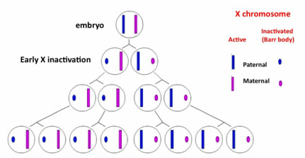
1) X chromosome inactivation (It may be useful for this section to refer to the karyotype figure on my homepage): In females, one of the two X chromosomes in each cell has to be "inactivated" (and in this process, once again, specifically modified histones and other epigenetic factors are involved :-) so only one of them is "expressed" in each cell. This process is a form of what is known as "dosage compensation" and it happens in many animal species to keep the amount of proteins made from X chromosomes the same in males and females. Genetically, one X chromosome in each female embryo came from the mother, and the other one from the father, so X inactivation results in either of these being inactivated in different cells. In males, the sex chromosome pair consists of one X (coming from the mother) and one Y (coming from the father), thus there is no need for X inactivation. X inactivation in females occurs early in development and in a random fashion. The inactive X chromosome suffers "condensation" and is visible under the microscope as a very dense smaller mass called a "Barr body". To put it simply, this condensation into a Barr body makes the genes in the inactive X chromosome inaccessible to proteins that would otherwise be responsible for these genes' expression into active proteins. Once a particular X chromosome is inactivated, the same one will be inactivated in all its daughter cells. However, at the initial stages one cell and its neighbor may have opposite X chromosomes inactivated, resulting in a "mosaic".
http://www.nytimes.com/2013/09/17/science/dna-double-take.html?pagewanted=all&_r=0
Chimeric "clones" can arise at different stages of development in either men or women due to mutations in cells which give rise to groups of descendant cells containing these mutations, or as cancer tissues which themselves contain mutated sequences compared to healthy tissues in the same person. There are at least 2 additional ways in which (only) women can think of themselves as chimeras: a combination of different types of cells rather than just the one we started with as a zygote. Although a more precise term would be micro-chimeras, as the group of cells inside some tissues that are different is quite small compared to the rest. One of these two women-specific chimera processes happens to all women, and the second only to those that have been pregnant:
1) X chromosome inactivation (It may be useful for this section to refer to the karyotype figure on my homepage): In females, one of the two X chromosomes in each cell has to be "inactivated" (and in this process, once again, specifically modified histones and other epigenetic factors are involved :-) so only one of them is "expressed" in each cell. This process is a form of what is known as "dosage compensation" and it happens in many animal species to keep the amount of proteins made from X chromosomes the same in males and females. Genetically, one X chromosome in each female embryo came from the mother, and the other one from the father, so X inactivation results in either of these being inactivated in different cells. In males, the sex chromosome pair consists of one X (coming from the mother) and one Y (coming from the father), thus there is no need for X inactivation. X inactivation in females occurs early in development and in a random fashion. The inactive X chromosome suffers "condensation" and is visible under the microscope as a very dense smaller mass called a "Barr body". To put it simply, this condensation into a Barr body makes the genes in the inactive X chromosome inaccessible to proteins that would otherwise be responsible for these genes' expression into active proteins. Once a particular X chromosome is inactivated, the same one will be inactivated in all its daughter cells. However, at the initial stages one cell and its neighbor may have opposite X chromosomes inactivated, resulting in a "mosaic".
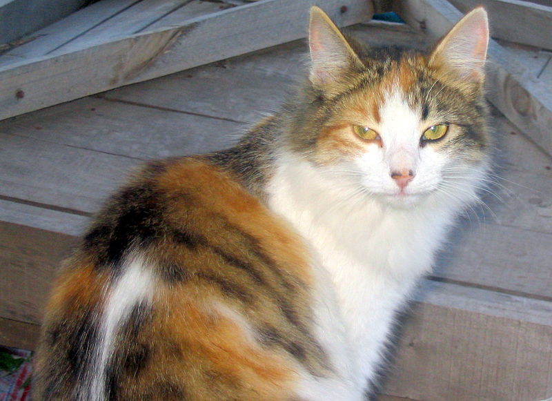
A classic visual example of these mosaics are female cats that are heterozygous for a coat color gene located on the X chromosome (X-linked). Heterozygote means the gene sequences on each of the two chromosomes in the pair (in this case, XX) are not the same, there are 2 different "alleles", one coming from the mother (located on the maternal chromosome X) and the other from the father (on the paternal X chromosome). This coat gene encodes a protein which results in orange hair color when there is one of these alleles present, or non-color when the alternative allele is (in which case the color may be black or white or a variation depending on additional genes located on other chromosomes that we are not focusing on here). Females with one allele for orange color and one for non-orange are tortoise shell and calico cats, visual mosaics of orange and other colors all over their coats. Each blotch contains only cells coming from a single cell in the embryo after X inactivation. For all of you cat lovers out there (myself included) you can now spot a female cat when you see an orange mosaic such as the one in the picture below (from wikipedia). Males will never be orange mosaics- they will show either all orange or no orange in their coats.
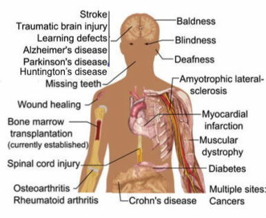
2) Mother incorporating fetal cells into her own tissues: We are all more or less aware of the huge potential that "stem cells" therapies offer for possible treatment of many diseases (including the ones shown in the figure below). In general, doctors try to go with "autologous" stem cells, cells from the same individual that needs the treatment. These stem cells can be "induced" to become a specific type of tissue after they are extracted from the donor (autologous or not), treated in the laboratory and then delivered into the donor's body.
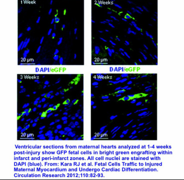
Fetal growth inside a woman's body offers possible stem cells circulating in the blood and resulting in fetal-maternal transfer into different tissues. Fetal blood contains a variety of stem cell types and during pregnancy; these "progenitor" cells circulate within maternal blood. Stem or progenitor cells have the capability of originating specific tissue cell types including blood, skin, liver and even heart. Fetal cells with this regenerative potential have been found in brain, liver, kidney, and lung injuries and are called fetal microchimeric cells. Initially, the big question was whether these microchimeric cells (fetal in origin but later on integrating into maternal tissues) are related to causing the injury or alternatively, they are targeted there to help the repair process by generating new healthy tissue where needed. Women with pregnancy-associated heart failure recover better compared to others with the same condition. In a study conducted in mice using an engineered GFP protein to mark fetal cells (see GFP section on my homepage), heart attacks were induced in pregnant mice and two weeks later, fetal cells were shown as 2% of the maternal heart and even formed blood vessels (see bright green GFP parts in the microscopy figure below from this study).
So....... the mother-fetus relationship may be more "symbiotic" (each of the two inside one body actually providing necessary "nutrients" or cells for the other's survival) than "parasitic" (only the mother feeding and providing for the fetus) in nature :-)

 RSS Feed